A comprehensive overview of the most mobile joint in our body, the shoulder, and the ways it can come unstuck.
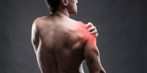
Pain in the shoulder is one of the more common conditions physiotherapists teat. The shoulder is the most mobile joint in the body and requires very complex muscular stability to maintain its function. The type of pain that you may be experiencing may give some indication as to the type of problem you are suffering from.
Shoulder pain is primarily felt in the upper arm either at the front or the outside of the shoulder and is associated with movements such as lifting your arm over-head, reaching out to the side, or reaching your hand behind your back, such as tucking in your shirt or doing up your bra.
The top 8 conditions related to the shoulder are:
- Subacrominal bursitis causing impingement
- Rotator cuff tendinopathy
- Rotator cuff tear
- Frozen shoulder
- Labral pathology – SLAP lesion
- Long head Biceps Tendinopathy
- Shoulder dislocation
- Arthritis
To better understand each of these conditions read on.
Shoulder Impingement syndrome
Just to be confusing, shoulder impingement syndrome has many names. It is also called subacromial impingement, painful arc syndrome, supraspinatus syndrome, swimmer’s shoulder, and thrower’s shoulder. It is a clinical syndrome which occurs when the sub-acromial bursa and tendons of the rotator cuff muscles become irritated and possibly inflamed as they pass through the subacromial space, resulting in pain and weakness at the shoulder.
Impingement syndrome is a very common condition that can be the result of overuse or isolated trauma such as a fall on to an outstretched hand or shoulder. Sometimes the pain which can be variable in nature is felt through an arc of movement. That is as you raise your arm up overhead the pain may only present through the middle portion of the movement. Generally, pain is only felt when moving the arm, and is not usually trouble at night unless you sleep of the sore side. In the absence of a fall the condition can take months to develop and some people can suffer from the condition for years.
Impingement syndrome is very treatable, with a high level of success with conservative treatment. The treatment approach at Physiodynamics is anchored in our ability to accurately diagnose the exact problem and mechanical dysfunctions that you are experiencing. A combined therapy of manual “hands-on” techniques and specialised exercises are essential if any conservative approach is to be successful.
At Physiodynamics, we can advise on the need / benefits and risks of certain medical approaches, such as steroid injections, as well as whether your condition requires further investigations or referral on to a specialist shoulder surgeon. We can explain your images such as X-rays, ultrasounds and MRI’s, in a way that you will understand.
Rotator Cuff Tendinopathy
The rotator cuff is a group of 4 muscles that are designed in such a way that they offer the shoulder stability through its amazing range of movement. These muscles act almost like a dynamic ligament holding the bones of the upper arm (humerus) and shoulder blade (scapula) together.
Without a functional rotator cuff you cannot place any force through the shoulder and you will lose range of movement and function, particularly over shoulder height. The rotator cuff tendons can become painful usually through over-use, and their symptoms are almost identical to that of bursitis. It is only through the application of specialised examination techniques that we can differentiate these two conditions.
If you have already seen a GP, and you have tried anti-inflammatory medications or even had a cortisone injection into the bursa with no relief of your symptoms, it is quite possible that your pain is associated to tendon pathology and pain. When we talk about tendinopathy, we are effectively describing a problem with the tendon. It used to be thought that it was swollen and inflamed, but recent research has proved otherwise. It is this reason that the more traditional medications such as anti-inflammatories and steroids are of little help.
We have found that a well-designed and specialised exercise program is essential in the successful management of a rotator cuff pathology. We don’t just look at this group of muscles. We must also consider the vast array of muscles that stabilise the shoulder blade as well. Furthermore, we find that complaints of this type adversely affect the function of the neck and upper-back, causing more pain and further muscular dysfunction. Our treatment approach therefore, must consider any and all of these structures as being involved in your problem.
Rotator cuff tear
Rotator cuff tears can occur because of one of two mechanisms. Firstly, and quite obviously, a traumatic injury that places a load upon the muscle and tendon that quite frankly is too great, and so often results in failure of the tendon and subsequent tearing.
Such mechanisms of injury might include falling and landing on the shoulder or arm or trying to prevent an object from falling from overhead. The heavier the object the greater the risk, but there are other factors that may influence the severity of the injury, such as health and age. The second common mechanism of tendon tears is that of a wear and tear nature. Repetitive tasks may lead to repeated trauma and failure of the tendons. This situation is more common in older populations, and in many circumstances these tears can occur without any pain and go un-noticed for years.
How do you know if you have a tear? Obviously if you have suffered a recent trauma to the arm or shoulder and are finding it difficult to or are unable to raise your arm above shoulder height, it is quite possible that you have a tear of a rotator cuff muscle. In many cases an accurate diagnosis can be made without the need of expensive radiology such as MRI’s.
Frozen (stiff) Shoulder
Adhesive capsulitis or ‘frozen shoulder’ is a disorder in which there is inflammation in the capsule that surrounds the shoulder (gleno-humeral) joint. This results in adhesions (i.e. unnatural connections) within the joint and contraction or shrinkage of this ‘capsule’.
There are 2 basic types of Frozen shoulder:
Primary or Idiopathic (of unknown cause) Frozen shoulder. This may occur spontaneously and without any precipitating factors.
Secondary Frozen shoulder occurs because of a known cause such as shoulder surgery or trauma.
Adhesive capsulitis results in limitation of both active and passive movement of the shoulder, causes pain at the extremes of motion, and interferes with normal daily activities. Unlike other shoulder problems, night pain may be severe. There may be a sense of restriction of the joint when it is passively moved. There is no way of telling who will develop a frozen shoulder, however, we do know that both men and women who suffer from diabetes are more at risk of developing both primary and secondary frozen shoulders. Accurate diagnosis of this condition is paramount. This condition can quite often be mis-diagnosed as something else and as such lead to unnecessary interventions’, pain and cost.
Labral Pathology
The labrum is a thin piece of triangular cartilage that helps the head of the humerus (ball) stay seated in the glenoid fossa (socket) of the scapula. Basically, it deepens the socket of the shoulder joint. This labrum is very important in helping to stabilise the shoulder joint, and as such some types of damage to it can result in quite severe dysfunction of the shoulder. The 2 most common labral injuries are SLAP lesions, and Bankart Lesions.
SLAP tears. The term SLAP stands for Superior Labrum Anterior and Posterior. In a SLAP tear the top part of the labrum (superior) is injured in the region of the Biceps tendon attachment. A SLAP tear occurs both in front of (anterior) and behind (posterior) to this attachment.
A SLAP tear may occur from traumatic injury such as falling onto an outstretched hand which may cause the labrum to be pinched, or rapid traction of the arm (say if you were to slip on a ladder and catch yourself). Overuse injuries associated with large amounts of a repetitive overhead action, such as packing shelves, lifting boxes overhead or throwing a ball.
Symptoms of a SLAP tear can be;
- Deep, aching pain.
- Popping, clicking, catching, locking or grinding in the shoulder.
- Decreased range of motion.
- Pain when lifting or carrying objects.
- Pain when moving the arm or shoulder.
- Decreased shoulder strength
Long head Biceps tendinopathy
The long head of the biceps tendon passes over the head of the humerus and crosses into the shoulder joint. The tendon can become irritated as a result of either rotator cuff pathologies and / or SLAP lesions. Differentiating the long head of biceps from these other pathologies can be quite difficult, due to the similarities in pain patterns and aggravating factors.
Patients with biceps tendinopathy may complain of a deep, throbbing ache in the anterior shoulder. Repetitive overhead motion of the arm initiates or exacerbates the pain.
Acromioclavicular joint pathology
The acromioclavicular (AC) joint is the connection between the acromion process of the scapula and the lateral end of the clavicle (collar bone). A joint is held in place by its capsule and the trapezoid and conoid coracoclavicular (CC) ligaments that connect the coracoid process of the scapula to the clavicle. The joint contains a fibrocartilaginous disc that cushions the articulations.
Two main conditions that affect this joint are dislocations and arthritis. AC dislocations are predominantly the result of sporting trauma. A fall onto the point of the shoulder or a full contact tackle in AFL, Rugby league, Rugby union are often the cause of the AC joint being dislocated. AC dislocations are quite easy to diagnose. They are characterised by a prominent elevation of the lateral end of the clavicle when compared to the un-injured side. An x-ray to determine the severity of the dislocation and to rule out any fracture is recommended.
In non-surgical cases, a structured rehabilitation program will restore your shoulder range of movement and strength. Return to activity is dependent upon pain and shoulder function.
AC arthritis, is a condition where the fibrous disc becomes worn and the end of the clavicle begins to rub against the acromion. This results in pain in the region which is quite difficult to isolate. The pain associated with AC arthritis can feel very similar to that of SLAP lesions and long head biceps tendinopathies. It is because of these complex interactions that we commonly see clients with long standing pain having been through multiple therapists and medical professionals with little success. Accurate diagnosis is essential and can only be achieved through a thorough assessment and elimination process.
Shoulder Dislocation
Shoulder dislocations are an extremely painful injury. Most dislocations happen at the lower front of the shoulder and are usually related to a trauma that sees the arm abducted and forcibly externally rotated. This usually occurs during a fall. The humerus moves forwards and downwards through the weakest part of the shoulder capsule and dislocates into a position in front of the shoulder blade. A less common dislocation is where a fall onto a straight arm may see the humerus dislocate posteriorly (backwards). This mechanism usually also results in a fracture of the shoulder socket.
In some circumstances a shoulder may become susceptible to dislocation through repeated overhead activities. Throwing sports can place repeated stress upon the capsule loosening it to a point that a dislocation is likely.
Causes and risk factors for shoulder dislocation include:
- Traumatic injury, such as a fall, sports injury, or other accident.
- Overuse/repetitive strain: from sports such as tennis, golf, swimming, volleyball
- Loose capsular ligaments: the connective tissue in the shoulder that keeps the head of the upper arm bone in the shoulder socket becomes loosened from injury or overuse, or from previous shoulder dislocations
- Multi-directional instability: this is generally the result of a genetic hypermobility condition. People, with this instability often have a type of connective tissue which makes their ligaments ‘looser’. We sometimes refer to this as being “double-jointed”.
Once a shoulder has been dislocated, it is at risk of further dislocations. The risk of further dislocations is somewhat age dependent.
- In people under the age of 20, 70 -100% may experience a recurrence of their shoulder dislocation.
- In the 20-30 year-olds age, the risk is 70-80%
- Over 50’s have a risk of recurrence of 15-20%
Recurrent dislocations can result in earlier development of osteoarthritis of the shoulder, which can be quite painful and limiting.
Arthritis
Described simply, arthritis is swelling of the joint. The two main types of arthritis are Osteoarthritis (OA) and Rheumatoid arthritis (RA).
Osteoarthritis
Commonly associated with “wear-and-tear”, osteoarthritis is a condition that mechanical use of the joint wears down smooth outer covering (articular cartilage) of bone. As the cartilage wears away and the protective space between the bones decreases. The underlying bone may become damaged and inflamed. During movement, the bones of the joint rub against each other, causing pain. Generally, this type of arthritis is seen in older people, and is considered a normal function of movement and aging. However, there are circumstances that can lead to earlier than expected development of OA.
The joints most likely involved in the shoulder are where the humerus connects with the scapula (Glenohumeral joint) and the joint between the clavicle and the top of the scapula (Acromioclavicular joint).
Symptoms of arthritis are usually pain, swelling and loss of movement. Glenohumeral arthritis will limit the amount of shoulder movement, particularly with overhead activities. Acromioclavicular arthritis whilst very painful and somewhat more common, does not always limit the shoulder range in the same way. Furthermore, this type of condition can sometimes present similarly to other shoulder conditions, such as labral and rotator cuff pathologies, hence an accurate diagnosis is very important before treatment.
Whilst OA is considered a normal fact of life, it is more common in weight bearing joints such as the knee and ankle. In the shoulder there are a few risk factors that if ignored can lead to the development of OA.
- Unstable shoulder. Recurrent dislocations of the shoulder cause an acceleration of the damage of the articular cartilage and the development of OA.
- Rotator cuff tears, chronic cuff tears, particularly of the supraspinatus muscle can lead to an inability of the rotator cuff to hold the head of the humerus in the glenoid fossa (socket). The humerus can move upward and rub against the acromion. This can damage the cartilage surfaces of the bones, causing arthritis to develop.
The combination of a large rotator cuff tear and advanced arthritis can lead to severe pain and weakness, resulting in significant loss of shoulder function.
Rheumatoid Arthritis
Rheumatoid arthritis (RA) is a chronic disease that attacks multiple joints throughout the body. It is an autoimmune disease, which means that the immune system attacks its own tissues. In RA, the immune system attacks the joint capsule lining and lubricating fluid, resulting in destruction of the cartilage, bone and joint capsule. Rheumatoid arthritis is equally common in both joints of the shoulder. RA joints are very swollen, and painful (inflamed).
If you have any questions, please give us a call or make an appointment.